Makuladegeneration test. Amsler Chart to Test Your Sight 2020-01-15
Is There a Test for Macular Degeneration?

Similar symptoms with a very different etiology and different treatment can be caused by or or any other condition affecting the macula, such as. The first signs of Age-related Macular Degeneration are typically discovered by an eye doctor in an annual dilated eye exam. Difficulty seeing in low light You may notice that you have become , this is called photophobia. Clinical trials testing new treatments, interventions and tests as a means to prevent, detect, treat or manage this disease. The size of the blind spot can change, becoming larger as damage to the macula progresses.
Next
Amsler Grid

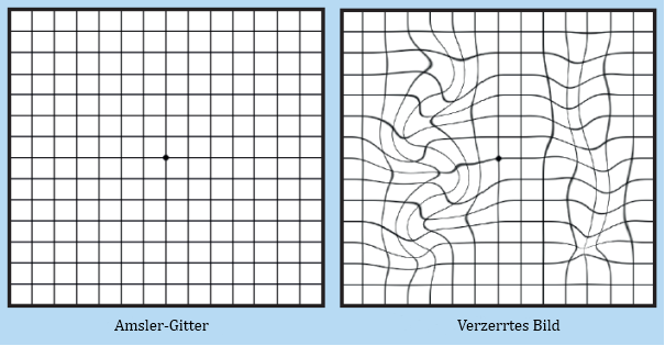
If normal retinal reception and image transmission are sometimes possible in a retina when high concentrations of drusen are present, then, even if drusen can be implicated in the loss of visual function, there must be at least one other factor that accounts for the loss of vision. Symptoms include, but are not limited to, an onset of blurry eyesight, a dimming of colors, straight lines appearing wavy, and blind spots in one's central field of vision. Macular degeneration Other names Age-related macular degeneration Picture of the showing intermediate age-related macular degeneration Symptoms or in the center of the Usual onset Older people Types Early, intermediate, late Causes Damage to the of the Genetics, smoking Prevention Exercising, eating well, not smoking Treatment injected into the eye, , Frequency 6. Omega 3 fatty acids for preventing or slowing the progression of age-related macular degeneration. Standard screening tests include the visual acuity exam the letter chart with an E at the top and an Amsler grid, which looks like graph paper.
Next
Macular degeneration

The Cochrane Database of Systematic Reviews. Also, the more damaged your macula is, the lower the likelihood of success. The Amsler grid is used to check whether lines look wavy or distorted, or whether areas of the visual field are missing. Now test your other eye. If new glasses don't help, ask for a referral to a low vision specialist.
Next
Macular degeneration

Mutations in this gene can also cause. Proceedings of the National Academy of Sciences of the United States of America. There is strong evidence that laser coagulation will result in the disappearance of drusen but does not affect. If you are experiencing any changes on the macular degeneration test, please call today to schedule an eye exam. After photodynamic therapy, you'll need to avoid direct sunlight and bright lights until the drug has cleared your body, which may take a few days. In 2004, Stone et al.
Next
Diagnosing Age

Any change from that is a signal that something may be awry. Yellow dye called fluorescein is injected into a vein, usually in your arm. But researchers are working at the level of the human genome, the code that tells the body how to build and rebuild itself. Early work demonstrated a family of immune mediators was plentiful in drusen. It is now a Medicare-covered service for patients enrolled in Medicare across the U.
Next
Diagnosing Age

Wear the glasses you normally wear when reading. You may also use a closed-circuit television system that uses a video camera to magnify reading material and project it on a video screen. At this point in time, genetic testing for Stargardt serves only two purposes: the first is for family planning; the second is to diagnose Stargardt in young people with symptoms of the disease. Awareness of macular degeneration is increasing as the baby boomer population continues to age. Urine and saliva are sometimes dark orange afterwards, a result of the body metabolizing the dye. If new glasses don't help, ask for a referral to a low vision specialist.
Next
AMDF

This noninvasive imaging test displays detailed cross-sectional images of the retina. Most of the advanced diagnostics for studying the presence or progression of macular degeneration involve making images of the fundus the inside back of the eyeball and the retina. During this test, your doctor injects a colored dye into a vein in your arm. You may need repeated treatments over time, as the treated blood vessels may reopen. Mitochondrial dysfunction may play a role. The risk of developing symptoms is higher when the drusen are large and numerous, and associated with the disturbance in the pigmented cell layer under the macula. Sridhar, an assistant professor of clinical ophthalmology at the Bascom Palmer Eye Institute of the University of Miami's Miller School of Medicine, the Amsler is more than a mere checkerboard.
Next
Macular degeneration

National Institute for Health and Care Excellence: Clinical Guidelines. This activates the verteporfin destroying the vessels. Structure and function of the eyes. So how do you find out if your eyes are at risk for developing the number one cause of legal blindness in seniors? Vitamin supplements For people with intermediate or advanced disease, taking a high-dose formulation of antioxidant vitamins and minerals may help reduce the risk of vision loss, the American Academy of Ophthalmology says. What you see at that visit is your baseline.
Next
Macular Degeneration Test

It is due to damage to the of the. Having your pupils dilated for the eye exam will affect your vision for a time afterward, so you may need someone to drive or accompany you after your appointment. In fact, the majority of people over age 60 have drusen with no adverse effects. During an eye exam, your eye doctor may use an Amsler grid to test for defects in your central vision. Early diagnosis can help preserve and save vision.
Next


