Syndesmose. Anatomy of the distal tibiofibular syndesmosis in adults: a pictorial essay with a multimodality approach. 2020-01-05
TightRope™ Fixation for Ankle Syndesmosis

Without stress radiographs, the tibiofibular diastasis is not identified. Main who understands the condition and is a willing surgeon to do something about it. The interosseous ligament lies between the tibia and fibula. This provides for potential advantages over screw fixation, including greater, more anatomic mobility of the joint, quicker return to weight bearing and sports, no osteolysis, and no need for hardware removal. Related Document: Causes Why do I have this problem? If pain is observed, a syndesmosis injury is suspected. In the setting of purely ligamentous injuries, the present authors have recently moved to a combination of screw and suture-button fixation.
Next
Optimal management of ankle syndesmosis injuries
If the syndesmosis is damaged, the ankle joint may become unstable. Return to full weight-bearing took 5. Traditional rehabilitation modalities are employed, including rest, ice, compression, and elevation, to reduce swelling and pain after surgery. X-rays should be obtained if an injury to the syndesmosis is suspected. On the initial visit, a general medical screen is required, consisting of questions concerning diabetes, vascular disease, neuropathy, alcohol use, and medication use.
Next
Squeeze Test
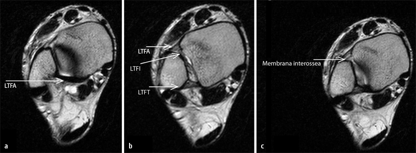
Protocols initially focus on controlling swelling and recovery from surgery. The intra-articularly injected green dye is visible in the tibiofibular recess 1 , which extends between the anterior 2 and posterior 3 tibiofibular ligament. Here are my 3-day post operative appointment photos of surgical sites, Click to enlarge photos First view of my leg 3-days after surgery Open reduction and internal fixation of Proximal Tibiofibular Syndesmosis with Arthrex TightRope at 3-day post -op appointment with my surgeon Dr. If a bimalleolar fracture occurs, screw fixation may be necessary to reduce the fibula. Patients report pain in varying degrees over the anterior and often posterior distal fibular joint.
Next
Distal tibiofibular syndesmosis

Since the deltoid ligament is essential for the stability of the ankle mortise, deltoid ligament repair helps to mitigate against future laxity. Kelly just wheeled me in the wheelchair across the parking lot to the restaurants. It can be used to repair a high ankle sprain, which damages the soft tissue structures between the tibia and fibula and causes these bones to separate. The fixation consisted of using 2 Arthrex Knotless TightRopes to dynamically stabilize the joint as well as a single bioabsorbable screw to statically stabilize the joint. Additionally, injuries can be more painful and take longer to heal than regular sprains, and high-impact activities, such as jogging and jumping, should generally be avoided until advised otherwise by a physician. It is based on these results that these authors uses a suture-button device to reduce syndesmosis injuries as well as Weber C fractures. This may involve operative or nonoperative management, followed by a structured rehabilitation program.
Next
Distal tibiofibular syndesmosis
Another test, called the squeeze test, is done by grabbing the calf just above the ankle joint and squeezing it. Initial range of motion exercises should be performed in the direction opposite that of the mechanism of injury to protect the ligament integrity. This plate is held in place by surgical screws. The Cotton Test assists you in identifying injury of the syndesmosis after a lateral ankle sprain. Due to the lack of flexibility in these joint structures, ligament injuries in these joints are not uncommon, particularly at the wrist and ankle.
Next
Distal tibiofibular syndesmosis
If a Maisonneuve fracture is present, fibular fixation is not performed for the proximal fibula fracture component. External rotation is not permitted within the first 6 weeks in the setting of a deltoid tear. The authors warn that they have not found the squeeze test to be as reliable when evaluating a syndesmosis injury. The ankle joint is a hinge joint. X-rays are usually taken every two weeks to make sure the ankle mortise isn't separating.
Next
TightRope™ Fixation for Ankle Syndesmosis

This instability, if uncorrected, can lead to chronic instability and significant morbidity, ultimately leading to degenerative arthritis. To begin the procedure, the surgeon bends the ankle slightly upward. The pain following this surgery was the most pain I have ever felt of any prior surgery along ; however, no pain then certainly no gain. Rehabilitation after surgery can be a slow process. A syndesmosis is defined as a fibrous joint in which two adjacent bones are linked by a strong membrane or ligaments. A careful progression to running and other impact activities begins a minimum of 12 weeks after surgery. They both had been in the operating room with Dr.
Next
Syndesmosis Ankle Ligament Injury

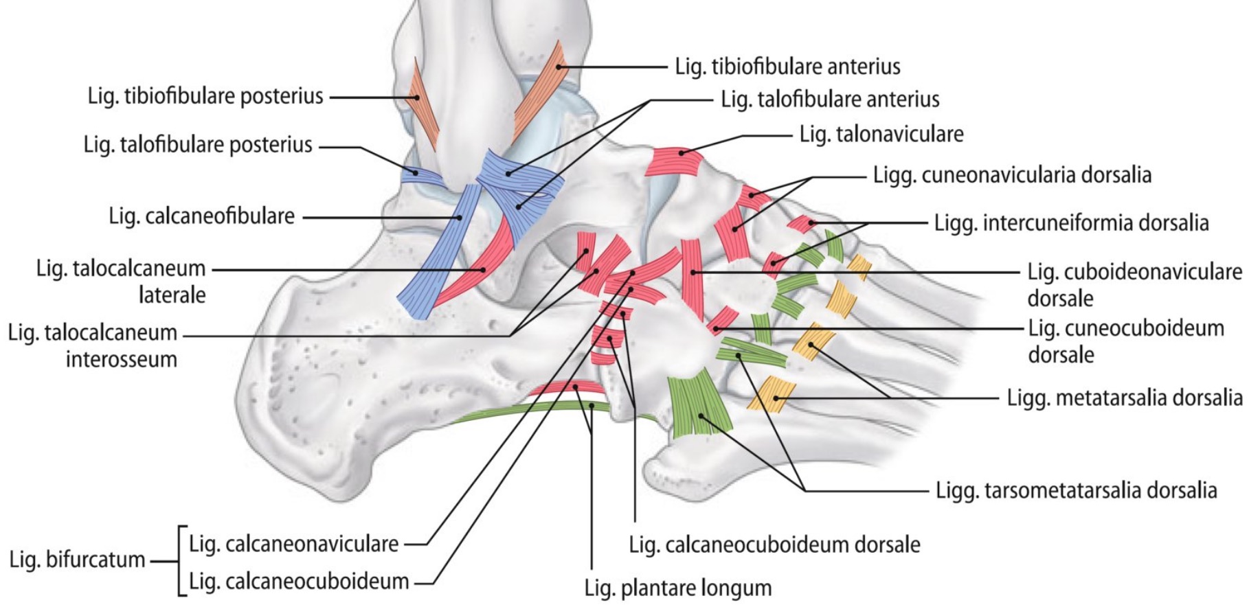
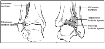
It may not be complete or timely. In doing research for all my surgical options, I also came across this published Elsevier Case Report research article: My story starts with the following video in the hotel room in the morning before leaving for surgery. X-rays are used to determine the severity of the syndesmosis injury. Dorsiflexion and eversion are also avoided until the ligament is healed. The distal tibia and fibula form the osseous part of the syndesmosis and are linked by the distal anterior tibiofibular ligament, the distal posterior tibiofibular ligament, the transverse ligament and the interosseous ligament. A gomphosis is a specialized fibrous joint in which a conical process or peg of one bone fits into a hole or socket in another bone.
Next
What does syndesmosis mean?
Besides feeling so good to give a loving touch to my entire body that just went through major stress, I strongly believe a massage is absolutely important to help the body detox from all the anesthesia and drugs given. Although the syndesmosis is a joint, in the literature the term syndesmotic injury is used to describe injury of the syndesmotic ligaments. After four weeks, patients are placed in a walking boot and allowed to gradually place more weight on their foot over another three to four weeks. These injuries occur commonly up to 18% of ankle sprains , and the incidence increases in the setting of athletic activity. The ankle joint is held in proper alignment by the ends of the tibia and fibula, which wraps around the inner and outer side of the ankle. The graded system classifies ankle instability according to mechanism of injury, ankle instability, and degree of ligament disruption.
Next
Fibrous joint

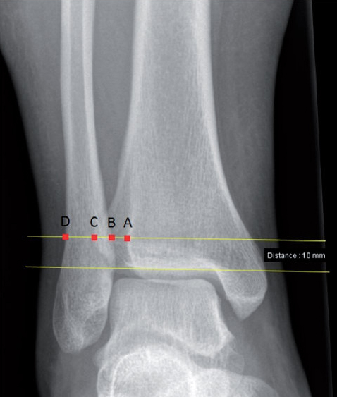
Additionally, a lateral talar displacement greater than 2 mm may result in greater than 90% chance of degenerative joint changes if left unaddressed because of increased pressure with decreased contact area. To perform an external rotation test, the patient is seated with the hips and knees flexed to 90°. I did not feel like I needed any pain medication. I had no problem getting and maintaining my full range of motion. Main, and they each reassured me that all had gone well.
Next