Bulbus caroticus. Bulbus Caroticus 2019-12-07
A. carotis interna

If the process is further distorted, the aorta lies predominantly over the right ventricle and the term double-outlet right ventricle is applied Figure 46-7. Because visceral obesity is a hallmark of insulin resistance and type 2 diabetes, we hypothesized that the association between arterial stiffness and visceral fat may be mediated by circulating inflammatory markers in patients with uncomplicated type 2 diabetes. Penderita Stroke saat ini menjadi penghuni terbanyak di bangsal atau ruangan pada hampir semua pelayanan rawat inap penderita penyakit syaraf. Eur J Cardiovasc Prev Rehabil 2010; 17: 18—23. Finally, endothelial function does not always correctly predict long-term outcome. Each vein receives the vertebral, deep cervical, deep thyroid, and internal thoracic veins.
Next
Die Plaquefläche an der Arteria carotis: Ein besseres Tool für die Risikostratifizierung in der Primärprävention als der PROCAM
This section does not any. Several cell types, including activated macrophages, lymphocytes, and smooth muscle cells, as well as adipose cells produce proinflammatory cytokines ,. The atria develop on the left and right sides of the heart and are thus truly lateralized structures. As the embryo develops, the primitive heart is displaced from the cephalic region into the thorax. Die neurologischen Ausfälle haben sich vollständig zurückgebildet. Vena mempunyai klep di sepanjang pembuluh darah.
Next
Carotid Sinus Massage
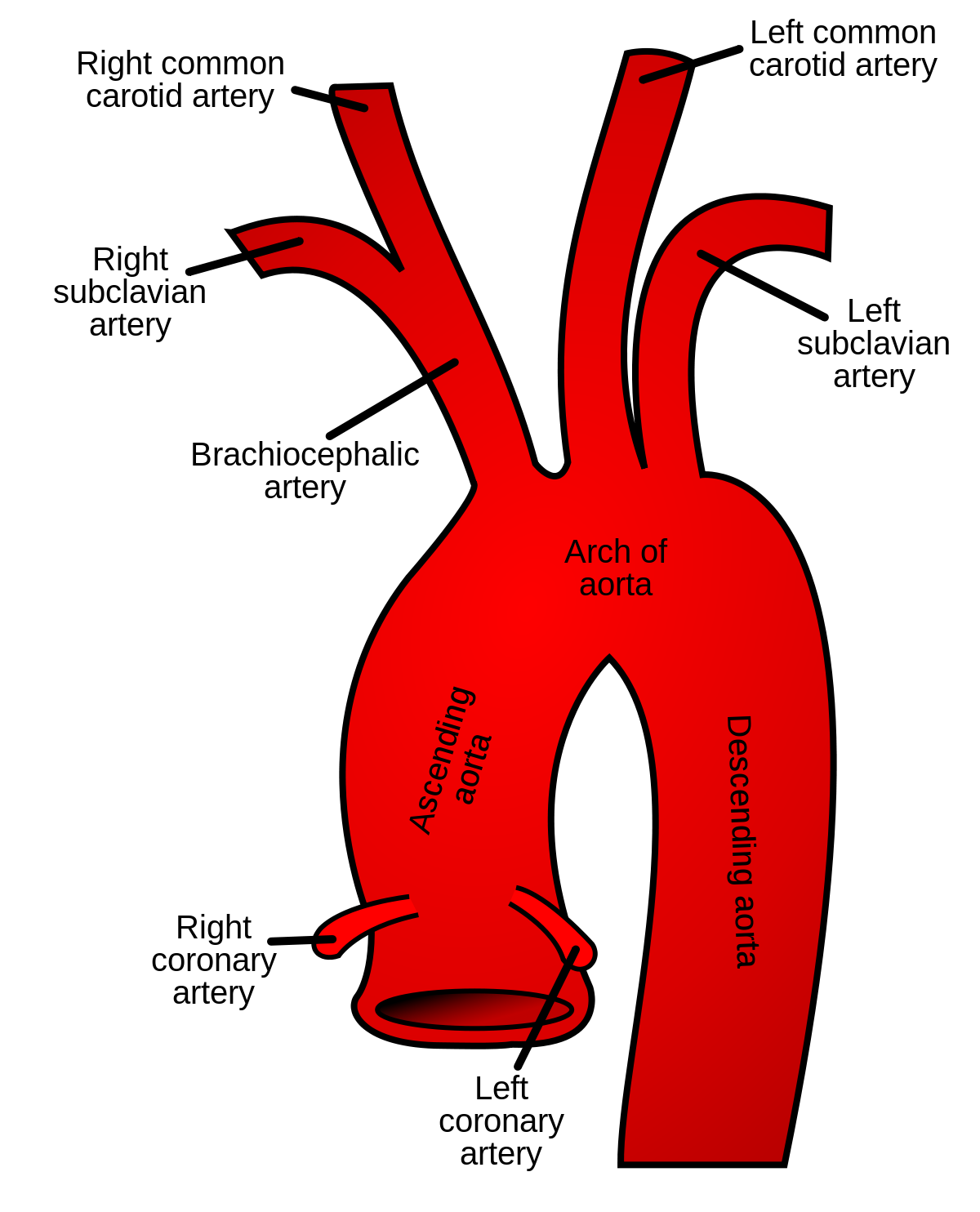
Indeed, the branching patterns of the right and left venae cavae may vary. Loss of Egfl7 function in zebrafish embryos specifically blocks vascular tubulogenesis. Brain metastasis: Unique challenges and open opportunities. Lin Clinical Breast Cancer 2019. Glomus tumors or malignant lesions of the skull base are a frequent cause of such lesions. There are two pairs of ridges bulbar and truncal that fuse to form a spiral septum that separates the aortic and pulmonary outflow tracts. This accounts for the partial spiraling of the aorta and the pulmonary artery in the adult heart.
Next
A. carotis interna

Die Messung bis zum hundertstel Millimeter ist nicht unproblematisch und die Dickenzunahme über die Zeitachse ist sehr gering. Lymphatic vasculature development is initiated by the specific expression of Prox1 in a subpopulation of vascular endothelial cells that subsequently adopt a lymphatic vasculature phenotype. A superior and an inferior vein originate from both an anterior and a posterior venous arcade. Functional inactivation of Prox1 in mice demonstrated that lymphangiogenesis requires the activity of this gene in a subpopulation of endothelial cells in embryonic veins. Der Cut off liegt bei den Männern für das Alter 35—44 Jahre bei 50 mm², 45—49 Jahre ab 70 mm², 50—54 Jahre ab 80 mm², 55—59 Jahre ab 90 mm², 60—64 Jahre ab 110 mm², bei den Frauen für das Alter 35—44 Jahre ab 30 mm², 45—49 Jahre ab 50 mm², 50—54 Jahre ab 70 mm² und 55—64 Jahre ab 90 mm².
Next
Bulbus cordis

Belcaro G, Nicalaides A, Ramaswami G et al. Additional genes implicated in abnormal atrial septal formation are listed in Table 46-2. The hepatic portal then receives a branch from the ventral abdominal vein before entering the liver. Blood from the hind limb may thus return to the heart through the posterior vena cava by several routes. Cite this article Liu, Z. Between them, the bulbus cordis extends from the ventricle anteriorly and slightly to the left.
Next
ardiana's: Artery Carotid Plaque
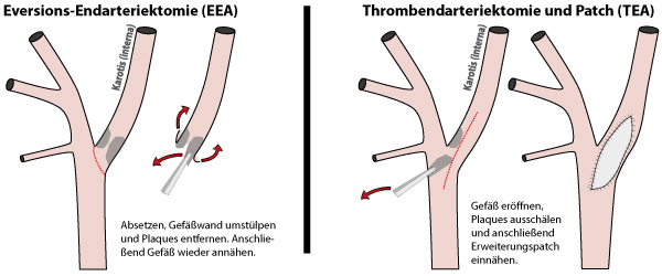
These venae cavae collect blood from the head and forelimbs, as well as the skin. They are largely a retrospective examination of clinical outcomes in patients enrolled in various research protocols in a few laboratories and hence may not be applicable to the population at large. Large artery stiffening involves structural changes, including fragmentation and degeneration of elastin, increases in collagen, and thickening of the vessel wall, but recent studies also suggest that endothelial dysfunction may play a pathophysiological role. The brachial artery followed a pattern similar to that of the femoral artery. Mengingat kecacatan yang ditimbulkan stroke permanen, sangatlah penting bagi usia muda untuk mengetahui informasi mengenai penyakit stroke, sehingga mereka dapat melaksanakan pola gaya hidup sehat agar terhindar dari penyakit stroke. High-resolution time-lapse 2-photon imaging of transgenic zebrafish was used to examine how endothelial tubes assemble in vivo, comparing our results with time-lapse imaging of human endothelial-cell tube formation in 3D collagen matrices in vitro.
Next
bulbus caroticus
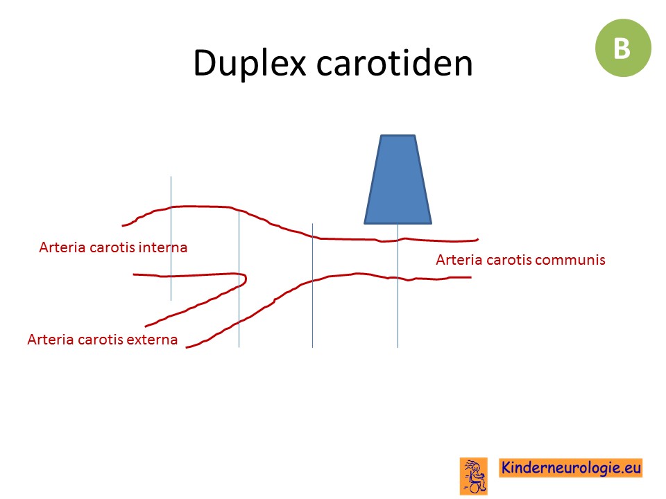
Ergebnisse: Bei 2361 gesunden Männern und 1011 Frauen im Alter zwischen 20 und 64 Jahren wurde mit Ultraschall die Fläche aller Plaques in der A. Subjects were examined in the supine position, with the head tilted slightly to the contralateral side. Bila ditinjau dari segi usia terjadi perubahan dimana stroke bukan hanya menyerang usia tua tapi juga menyerang usia muda yang masih produktif. Rhythmic changes in intrathoracic pressure during and peripheral increases in interstitial pressure of compression of lymph vessels, for example, due to , propel the lymph through the thoracic duct into the left subclavian vein lymph from avoce the diaphragm returns to the circulation through the bronchomediastinal duct or the right lymph trunk. Ein weiterer Nachteil ist, dass nur ein kleiner Teil der A.
Next
Bulbus cordis

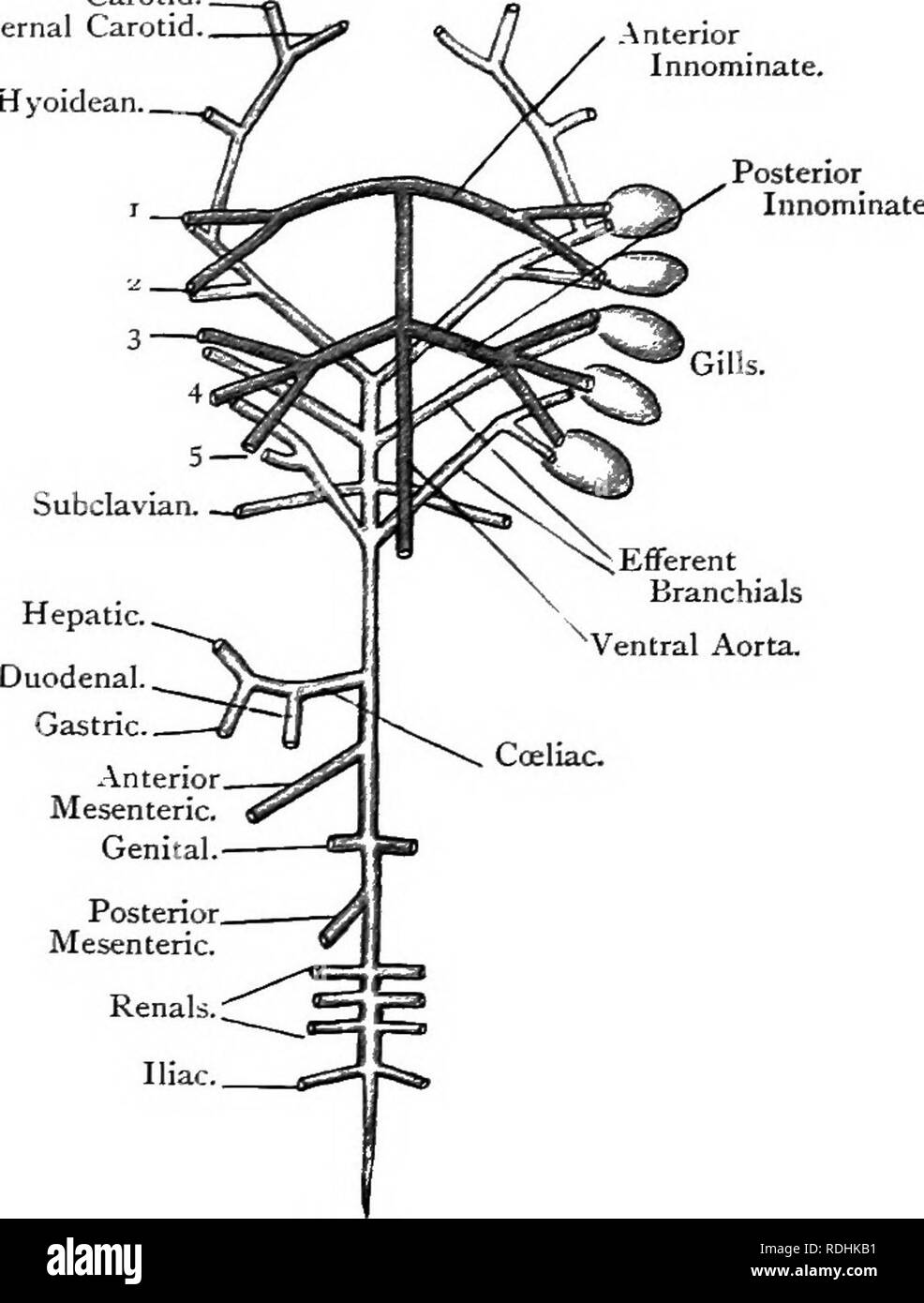
It supplies most of the rest of the body with blood, and so is the largest of the three branches. The ventral pharyngeal arteries, which are the precursors of the definitive external carotid arteries, branch directly from the ventral aortic sac and initially contribute to supply of the first and second aortic arches. Angka kejadian stroke dunia diperkirakan 200 per 100. The renal portal vein passes anteriorly, sending numerous branches into the kidney. This diagram depicts how the primitive heart pumps blood into the primitive embryonic circulation, which is constituted by a pair of cephalic dorsal aortae that are fused more caudally into a single descending aorta. The posterior vena cava also receives blood from much of the hind limbs and dorsal body musculature by way of the paired renal portal veins, which enter the kidneys Figures 6.
Next
ardiana's: Artery Carotid Plaque

Pembuluh ini berfungsi sebagai distributor zat-zat penting ke jaringan yang memungkinkan berjalannya berbagai proses dalam tubuh. Interessenkonflikt: Der Autor erklärt, dass er in keinem Interessenkonflikt steht. It is formed mainly by the following vessels: the gastric vein, which collects several vessels and drains the stomach and part of the esophagus, and the intestinal vein, formed by vessels that drain most of the small intestine and large intestine Figures 6. Perzentil der 35- bis 39-jährigen Männer und Frauen bzw. Right left lateral view ; d ventricle; e atrioventricular canal; f early left atrium; g sinus venosus; h truncus arteriosus; i cardinal vein inflow to sinus venosus.
Next
bulbus urethra
The anterior ciliary veins follow the anterior ciliary arteries, and receive branches from the sinus venosus, sclerae, the episcleral veins, and the tunica conjunctiva bulbi. Emerging strategies for treating brain metastases from breast cancer. By week 3 in the humans, cells from the first heart field coalesce along the ventral midline to form the primitive heart tube. Right left lateral view : e truncus arteriosus; f bulbus cordis; g ventricle; h atrioventricular canal; i early left atrium. The septum secundum grows adjacent to the septum primum as a thick muscular fold.
Next


